Film Diagnosis and treatment of lung cancer.. (1986)
Part 1
0:09:58
Part 2
0:09:42Diagnosis and treatment of lung cancer. (1986)
Annotation:
Educational film for students medvuzov.
Reel №1


Discussion of diagnoses of patients at an extended consultation at the cancer center.
The patient tells about his feelings.
A plane takes off, polluted air is around it.
A city covered in smog.
Smoking pipes.
Exhaust pipes of cars.
Map of the USSR, where the places with the highest incidence of lung cancer are marked.
The increase in the number of lung cancer diseases in numerical terms.
City polyclinic.
Preventive inspection.
Interviewing a visitor at the remote control of a minicomputer.
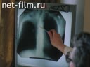
The doctor examines the X-rays of the lung.
For the high-risk group and for men over 50 years of age, pictures are taken on inhalation and exhalation and in lateral projections.
The data obtained are compared for the previous 3 years.
This technique allows timely detection of focal round shadows and violations of bronchial patency.
Dispensary examination.
Interview of the patient.
The doctor listens to the patient's lungs with a stethoscope.
The symptoms of lung cancer are not specific, gradually increasing cough, mucous and mucopurulent sputum, fever, chest pain.
All these are signs of the disease and they should be evaluated together.
The method of examination of a patient with suspected lung cancer.
Lung X-ray.
According to the results of the fluorography, the attending physician reports the diagnosis to a consultation of doctors.
Based on the fluographic images, graphic drawings of lung cancer development variants have been compiled.
When a diagnosis of lung cancer is established, patients are sent to specialized oncological institutions for further examination and the choice of a treatment method.
The diagnosis of lung cancer must be confirmed by morphological examination of the material obtained during bronchoscopy.
It can also visually confirm the nature of the changes and the prevalence of the process in the bronchi.
The material for further study can be obtained with controlled bronchial catheterization.
Under the control of the X-ray screen, a scraping is taken for cytological examination using a special brush.
Forms of lung cancer detected by morphological examination.
Cartoon explaining lymphogenic metastasis, hematogenic metastasis and the localization of metastases.
Key words
Lung cancer.
Metastases.
Medical examination.
Surveys.
Reel №2


The TNM system.
T1 - indicates the size of the tumor up to 3 cm.
T2 - more than 3cm .
T3 indicates the transition of the neoplasm to neighboring anatomical structures.
N1 means intrapulmonary and root metastases.
N2 - mediastinal metastases.
M indicates distant metastases.
The classification is accompanied by examples on X-rays.
The classification of the disease can be judged by the study on an X-ray computed tomograph.
On the screen of the tomograph, you can get a cross-section of any organ with an interval of up to 2 mm.
Episode of the discussion of the diagnosis at the doctors' consultation.
The patient is being prepared for surgery.
Surgery to remove the affected part of the bronchi and lung.
Cartoon explaining the operation of lung lobectomy and pneumonectomy.
It is very difficult to establish a correct diagnosis.
Even an experienced radiologist cannot always tell whether it is a neoplasm or a tuberculoma.
Unfortunately, percutaneous puncture of a neoplasm, under the control of an X-ray, also does not always give an informative answer.
Then diagnostic thoracotamia is indicated.
If a benign tumor is detected during the lung revision, then a precision tumor removal technique is used that spares the lung tissue as much as possible.
With a locally widespread process and the impossibility of surgical treatment, radiation therapy is indicated.
Interview of the patient by doctors.
Radiation treatment is impossible with copious hemoptysis, tumor decay, purulent inflammatory process in the lung.
The report of the doctor on the diagnosis of the interviewed patient.
If distant metastases are detected, the patient is prescribed chemotherapy.
Foci of increased accumulation of technetium 99 in bone metastases are shown.
Medications used in chemotherapy.
In some cases, not only the full effect is achieved, but also a long-term remission.
The visitor of the polyclinic answers the questions of the mini-computer questionnaire in the polyclinic.
The success of lung cancer treatment in early diagnosis.
Key words
Radical treatment of lung cancer.
Radiation therapy.
Chemotherapy.